2d Colour Doppler Echocardiography

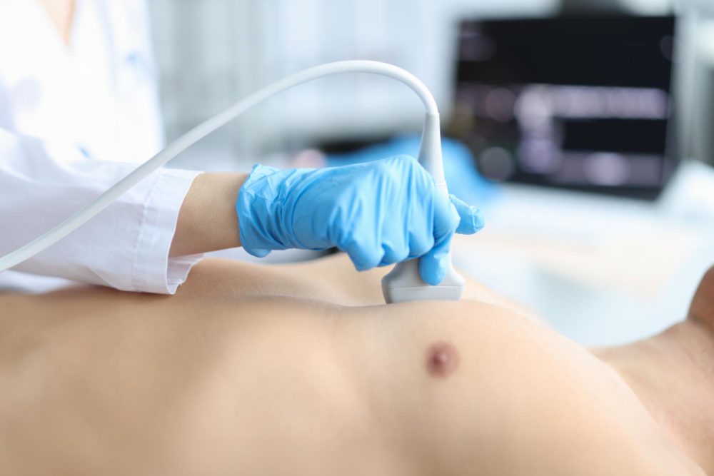
An Echocardiogram uses ultrasound to visualize your heart, its rhythm, and blood pumping. It’s useful for diagnosing valve or chamber issues.

A Contrast Echocardiogram enhances the view by injecting a special agent into your arm vein, improving the visibility of heart structures on ultrasound images.

An echocardiogram (echo) is an ultrasound scan of the heart that uses sound waves to create live images showing the structure and pumping function of your heart.

Why it’s used: It provides detailed information about how your heart chambers and valves are working, assesses heart muscle strength, discovers valve disease, detects cardiomyopathies, and monitors changes in heart function over time

How long it takes: The test typically takes about 30–45 minutes for a standard transthoracic echo

How to prepare: For most standard echoes, no special preparation is required — you may eat and drink normally and take your usual medications.

Wear comfortable clothes and remove any upper-body jewelry. If the echo is a more specialised type (stress echo or transesophageal echo), your doctor may provide specific instructions (e.g., avoid food, caffeine, or alcohol).

What to expect: You’ll lie on your left side while a technologist applies a gel on your chest and moves a small probe to capture images of your heart in motion. You may be asked to hold your breath or change positions briefly. After the scan, you will wipe off the gel and you’re free to go.

Results & follow-up: A cardiologist reviews the images and sends a report to your referring doctor, who will discuss the findings and next steps with you.

Scroll to Top